|
|
|
A number of scientific articles indicate that some of the alterations in cell signaling, which occur in cancer progression, are mediated by lipid second messengers and that most of these molecular signals are the immediate products of lipid metabolism. For instance, sphingosine kinase1 and 2 (SK1 and SK2) phosphorylates sphingosine to generate sphingosine 1-phosphate (S1P) which can be reversibly dephosphorylated by sphingosine phosphatase1 and 2 or irreversibly degraded by a pyridoxyl-phosphate-dependent S1P lyase. SK1 is thought to be oncogenic and based on recent animal studies developed inhibitors of SK1 have shown great potential as effective chemotherapeutic agents. Equally important, SK2 is involved in the immune response and compounds targeted at extracellular S1P signaling are demonstrating great promise in clinical trials for autoimmune diseases. Given that both the substrate and the product of the above enzymes are potentially important signaling molecules, the development of highly sensitive analytical techniques to monitor the enzyme activity and quantify the substrates and products of these enzymatic reactions either in a single cell or in a large cell population is extremely important.
Conventionally, tissues or cells of interest are extracted, and cell-free extracts are purified to obtain enzymes, which are employed for in vitro activity measurements. With current gene expression technology, specific lipid kinase, phosphatase, or phospholipase isoforms have been cloned and expressed as recombinant proteins suitable for study of enzyme activity and discovery of new inhibitors. However, many assays still require radioisotopic labels which are expensive and require intricate procedures to acquire, and a labor intensive process to separate substrates and reagents from products before they can be detected. In addition, radiometric separation methods are poorly suited to high throughput screening; either for diagnosis or for drug discovery, and very few established methods for the characterization of lipid modifying enzymes provides adequate information on the subcellular distribution of enzyme activity.
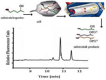 As stated earlier, our goal is to develop capillary electrophoresis (CE) separation methods that can be used to measure enzyme activity either in a large cell population or in single cells. As stated earlier, our goal is to develop capillary electrophoresis (CE) separation methods that can be used to measure enzyme activity either in a large cell population or in single cells.
The optmized CE separation methods using fluorescent lipids may find applications in drug screening as well as development of cell-based assays for a number of diseases including cancer.
Back to Research Page |
|